Navigating an anterior cruciate ligament injury often begins with a knee MRI to confirm the diagnosis. A sudden popping sound followed by intense pain can leave you wondering about the severity of the damage to your joint.
Understanding when medical professionals recommend advanced imaging can help you prepare for the road to recovery. In short…
- Accurate diagnosis matters. An MRI confirms the presence and severity of a ligament tear.
- Symptoms guide imaging. Doctors recommend scans based on specific signs like instability and swelling.
- Treatment depends on it. The detailed results from an MRI determine if you need surgery.
What is the Anterior Cruciate Ligament?
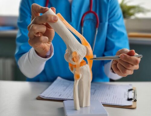
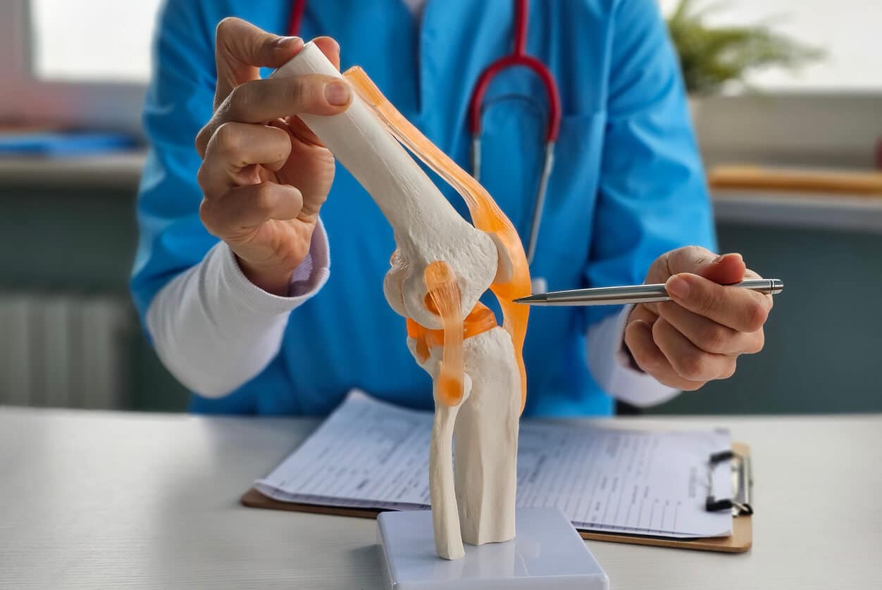
The anterior cruciate ligament (ACL) acts as one of the key stabilizers in your knee joint. It connects your thighbone (femur) to your shinbone (tibia) in the center of the knee. This ligament prevents the tibia from sliding out in front of the femur during movement.
Athletes often injure this ligament during sports that involve sudden stops or changes in direction. However, non-athletes can also suffer tears from falls or awkward landings. Understanding the anatomy helps explain why accurate diagnosis is so critical for future mobility.
What Are the Classic Signs of a Torn ACL?
Most patients report hearing or feeling a distinct “pop” at the moment of injury. This is often followed by significant swelling that occurs within a few hours.
Another hallmark sign involves the knee feeling unstable or “giving way” under your weight. You might also experience a loss in full range of motion. If you notice these symptoms, it is essential to seek medical attention immediately.
Does Every Knee Injury Require a Knee MRI?
Orthopaedic specialists typically begin with a physical examination before ordering an MRI. They may perform specific movements, such as the Lachman test, to check the laxity of the ligament.
While X-rays don’t show the ligament, they can sometimes reveal a ‘Segond fracture’—a small bone chip on the side of the knee that is a strong indicator of an ACL tear. However, X-rays do not show soft tissues like ligaments or tendons effectively. If the X-ray is negative but pain persists, further imaging becomes necessary.
When is a Knee MRI Recommended?
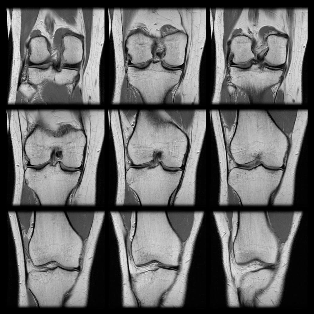
Your doctor will likely order a knee MRI if the physical exam suggests a tear but remains inconclusive. This scan uses strong magnetic fields and radio waves to create detailed images of the knee.
It allows physicians to see the extent of the rupture and any associated damage. An MRI is particularly useful when the knee is too swollen to be examined physically. It provides a definitive look at the soft tissue structures inside the joint.
Checking for Associated Injuries
ACL injuries rarely happen in isolation. An MRI helps identify concurrent injuries to the meniscus or other ligaments.
Identifying these issues early ensures that the surgical plan addresses all damaged structures. A comprehensive diagnosis leads to better long-term outcomes for the patient.
What Does Research Say About Imaging Accuracy?
The accuracy of magnetic resonance imaging makes it the gold standard for diagnosing soft tissue injuries. According to the Mayo Clinic, an MRI provides a much clearer image of soft tissues than other methods. This clarity allows for precise surgical planning when reconstruction is necessary.
A study highlighted that MRI scans are highly sensitive for detecting complete ACL tears. The researchers found that MRI offers crucial data that influences the decision between conservative management and surgical reconstruction.
Another study in the Archives of Bone and Joint Surgery confirmed that MRI helps avoid unnecessary diagnostic arthroscopy. This research emphasizes that non-invasive imaging should always precede invasive diagnostic procedures.
How Should You Prepare for a Knee MRI for an ACL Injury?
Undergoing a knee MRI is a painless and non-invasive procedure. You will need to remove all metal objects, including jewelry, watches, and glasses.
The technician will position your leg carefully to ensure clear images of the joint. The machine may make loud tapping or thumping noises during the scan. The entire process typically takes between 30 and 60 minutes.
Are There Alternatives to an MRI?
While a knee MRI is the preferred method, other options exist for specific situations. Ultrasound may visualize superficial ligaments, but cannot see deep inside the knee joint well.
Diagnostic arthroscopy involves inserting a tiny camera into the knee, but this is an invasive surgical procedure. Therefore, doctors almost always prefer an MRI as the primary diagnostic tool.
Frequently Asked Questions About Knee Diagnostics
Can a Physical Exam Miss an ACL Tear?
Yes, muscle guarding or severe swelling can make it difficult for a doctor to feel the ligament tear. This is why imaging is often required for confirmation.
Is Radiation Involved in an MRI?
No, unlike a CT scan or X-ray, an MRI does not use ionizing radiation. It is considered safe for most patients.
How Long Does It Take to Get Results?
A radiologist must review the images before sending a report to your orthopaedic doctor. This process usually takes a few days.
Getting a Knee MRI for an ACL Tear in Cary, NC
If you suspect a ligament injury, obtaining a timely knee MRI in Cary or the Triangle area is vital. Our board-certified specialists at Cary Orthopaedics provide comprehensive care for acute knee conditions.
We utilize advanced diagnostics to create personalized treatment plans for every patient. Whether you require physical therapy or minimally invasive surgery, we are here to help you restore your mobility.
Contact us today at (919) 467-4992 or use our appointment form to schedule an appointment.
